Brief Guide to the CTE Brains in the News. Part 2: Fred McNeill
Chronic traumatic encephalopathy (CTE) is the neurodegenerative disease of the moment, made famous by the violent and untimely deaths of many retired professional athletes. Repeated blows to the head sustained in contact sports such as boxing and American football can result in abnormal accumulations of tau protein (usually many years later). The autopsied brains from two of these individuals are shown below.
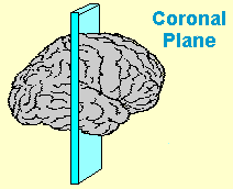
Left: courtesy of Dr. Ann McKee in NYT. Right: courtesy of Dr. Bennett Omalu in CNN. These are coronal sections1 from the autopsied brains of: (L) Aaron Hernandez, aged 27; and (R) Fred McNeill, aged 63.
Part 1 of this series looked at complicating factors in the life of Aaron Hernandez — PCP abuse, death by asphyxiation — that presumably had some impact on his brain beyond the effects of concussions in football.
Part 2 will discuss the tragic case of Fred McNeill, former star linebacker for the Minnesota Vikings. He died in 2015 from complications of Amyotrophic Lateral Sclerosis (ALS), suggesting that his was not a “pure” case of CTE, either.
Fred McNeill
McNeill in 1974 (Mike Zerby / Minneapolis Star Tribune).
Obituary: Standout of the 1970s and 1980s was suffering from dementia and died from complications from ALS, according to Matt Blair [close friend and former teammate]
ALS is a motor neuron disease that causes progressive wasting and death of neurons that control voluntary muscles of the limbs and ultimately the muscles that control breathing and swallowing. Around 30-50% of individuals with ALS show cognitive and behavioral impairments.
According to a recent review (Hobson and McDermott, 2016):
Overlap between ALS and other neurodegenerative diseases, in particular frontotemporal dementia (FTD) and parkinsonism, is increasingly recognized. ...
Approximately 10–15% of patients with ALS show signs of FTD ... typically behavioural variant of FTD. A further 50% experience mild cognitive or behavioural changes. Patients with executive dysfunction have a worse prognosis, and behavioural changes have a negative impact on carer quality of life.
This raises the issue that repetitive head trauma can result in multiple neurodegenerative diseases, not only CTE.2 In fact, this has been recognized by other researchers who studied 14 retired soccer players who were experts at heading the ball (Ling et al., 2017). Only four had pathologically confirmed CTE:
...concomitant pathologies included Alzheimer's disease (N = 6), TDP-43 (N = 6), cerebral amyloid angiopathy (N = 5), hippocampal sclerosis (N = 2), corticobasal degeneration (N = 1), dementia with Lewy bodies (N = 1), and vascular pathology (N = 1); and all would have contributed synergistically to the clinical manifestations. ... Alzheimer's disease and TDP-43 pathologies are common concomitant findings in CTE, both of which are increasingly considered as part of the CTE pathological entity in older individuals.
So the blanket term of “CTE” can include build-up of not only tau, but other abnormal proteins typically seen in Alzheimer's disease (Aβ) and the ALS-FTD spectrum (TDP-43). This lowers the utility of an in vivo marker specific to tau in diagnosing CTE in living individuals, an important enterprise because definitive diagnosis is only obtained post-mortem.
This brings us to the problematic report on Mr. McNeill's brain and the news coverage surrounding it.
CTE confirmed for 1st time in live person, according to exam of ex-NFL player
The recent study by Omalu and colleagues (2017) performed a PET scan on Mr. Neill almost 4.5 years before he died. This was before any motor signs of ALS had appeared. Clearly, 4.5 years is a very long time in the course of progressive neurodegenerative diseases, so right off the bat a comparison of his PET scan and post-mortem pathology is highly problematic.
Former Vikings linebacker Fred McNeill identified as subject of breakthrough CTE study
Another reason this study was not the “breakthrough” of news headlines is because the type of pathology plainly visible on MRI, and the type of cognitive deficits shown on neuropsychological tests, were quite typical of Alzheimer's disease and perhaps also vascular dementia. The MRI scan taken at the time of PET “showed mild, global brain atrophy with enlarged ventricles, moderate bilateral hippocampal atrophy, and diffuse white matter hyperintensities.”
Among his worst cognitive deficits at the time of testing were memory and picture naming, which is characteristic of Alzheimer's disease (AD). Likewise, the behavioral deficits reported by his wife are typically seen in AD.
Two years after the PET scan, he developed motor symptoms of ALS. His wife noted he could no longer tie his shoes or button his shirts. He developed muscle twitching in his arms and showed decreased muscle mass in his arms and shoulders. He was diagnosed with ALS 17 months prior to death, which was in addition to his presumed diagnosis of CTE.
FDA says no to marketing FDDNP for CTE
Finally, the molecular imaging probe used to identify abnormal tau protein in the living brain, [18F]-FDDNP, is not specific for tau. It also binds to beta-amyloid and a variety of other misfolded proteins. Or maybe not!
As I've written before, the brain diagnostics company TauMark™ was admonished by the FDA for making false claims. Six authors on the current paper hold a financial interest in the company. Most other research groups use more specific tau imaging tracers such as [18F]T807 (aka [18F]AV-1451 or Flortaucipir).
I certainly acknowledge that theses types of pre- and post-mortem studies are very difficult to conduct, and although the n=1 is a known weakness, you have to start somewhere. Nonetheless, the stats relating FDDNP binding to tau pathology were very thin and not all that believable. The paragraph below presents the results in their entirety. Note that p=.0202 was considered “highly correlated” while p=.1066 was not significant.
Correlation analysis was performed to investigate whether the in vivo regional [F-18]FDDNP binding level agreed with the density of tau pathology based on autopsy findings. Spearman rank-order correlation coefficient (rs) was calculated for the regional [F-18]FDDNP DVRs (Figure 1) and the density of tau pathology, as well as for amyloid and TDP-43 substrates (Table 5). Our results showed that the tau regional findings and densities obtained from antemortem [F-18]FDDNP-PET imaging and postmortem autopsy were highly correlated (rs = 0.592, P = .0202). However, no statistical correlation was found with the presence of amyloid deposition (r s = -0.481; P = .0695) or of TDP-43 (rs = 0.433; P = .1066).
Also, FDDNP-PET showed that in cortical regions, the medial temporal lobes showed the highest distribution volume ratio (DVR), along with anterior and posterior cingulate cortices. Isn't this typical of the Aβ distribution in AD?
I'm not denying the existence of CTE as a complex clinical entity, or saying that multiple concussions don't harm your brain. Along with others (e.g., Iverson et al., 2018), I'm merely suggesting that the clinical, cognitive, behavioral, and pathological sequelae of repeated head trauma should be carefully studied, and not presented in a sensationalistic manner.
Footnotes
1 Illustration of the coronal plane of section.

2 Note that most cases of ALS and FTD are not caused by concussions.
Read Part 1 of the series:
Brief Guide to the CTE Brains in the News. Part 1: Aaron Hernandez
References
Hobson EV, McDermott CJ. (2016). Supportive and symptomatic management of amyotrophic lateral sclerosis. Nat Rev Neurol. 12(9):526-38.
Iverson GL, Keene CD, Perry G, Castellani RJ. (2018). The Need to Separate ChronicTraumatic Encephalopathy Neuropathology from Clinical Features. J Alzheimers Dis. 61(1):17-28.
Ling H, Morris HR, Neal JW, Lees AJ, Hardy J, Holton JL, Revesz T, Williams DD. (2017). Mixed pathologies including chronic traumatic encephalopathy account fordementia in retired association football (soccer) players. Acta Neuropathol. 133(3):337-352.
Omalu B, Small GW, Bailes J, Ercoli LM, Merrill DA, Wong KP, Huang SC, Satyamurthy N, Hammers JL, Lee J, Fitzsimmons RP. (2017). Postmortem Autopsy-Confirmation of Antemortem [F-18] FDDNP-PET Scans in a Football Player With Chronic Traumatic Encephalopathy. Neurosurgery. 2017 Nov 10.
Further Reading – I've written about CTE a lot, you can read more below.
FDA says no to marketing FDDNP for CTE
Is CTE Detectable in Living NFL Players?
The Ethics of Public Diagnosis Using an Unvalidated Method
The Truth About Cognitive Impairment in Retired NFL Players
Lou Gehrig Probably Died of Lou Gehrig's Disease
Blast Wave Injury and Chronic Traumatic Encephalopathy: What's the Connection?
Little Evidence for a Direct Link between PTSD and Chronic Traumatic Encephalopathy
Subscribe to Post Comments [Atom]


0 Comments:
Post a Comment
<< Home